Three Radiological Technology students at The Michener Institute of Education at UHN recently had the unique opportunity to take X-ray images during a cadaver lab in the Temerty Advanced Surgical Education & Simulation Centre on Michener’s 12th floor.
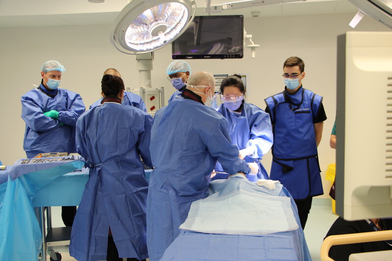
British multinational medical equipment manufacturing company Smith + Nephew (S&N) contacted the Temerty Centre about hosting an orthopedic ankle surgery course on October 28 using space and equipment on the 12th floor. As part of the course, S&N required X-ray imaging be taken throughout the procedure to check on the placement of the implant.
“We’re trying to find different ways to integrate Michener’s full-time programs into any type of activity we have happening within the Centre,” says Karen Chaiton, Director of Business Operations at University Health Network. “When we learned that imaging would be required for this lab, we thought it would be a perfect opportunity to pull in some Radiological Technology students and offer them a greater learning opportunity.”
“Being able to offer our S&N learners a training opportunity as close to real world as possible is critical to a successful learning experience and early adoption of new technology,” says Mark Hynes, Regional Sales Manager, Ontario East, S&N. “In addition to the aforementioned benefits to our learners, we welcome opportunities to have Michener students partake in our labs. Creating teaching environments for all professions that support our technology and procedures only helps to ensure successful and safe outcomes.”
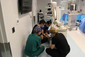
While several Radiological Technology students expressed interest in being part of the lab, second-year students Tian Hua, Stephanie Olson and Adam Magliozzi were randomly selected to participate. Associate Professor Desmond Chau was also present to guide the students on how to use the C-Arm X-ray machine, which is a medical imaging device with a C-shaped arm that can move around the body.
In their first year, the students only had virtual exposure to the C-Arm by watching their instructor demonstrate how to set up and use the machine over a live stream, so most of the second-year students in the Fall semester have not yet used the equipment clinically.
“This lab is very close to what they would actually be doing in an operating room and it also gave us a chance to talk about what happens in the periphery, such as how we set up, plan ahead, how we stay out of the way of the surgeons and maintain a sterile field,” says Desmond Chau.
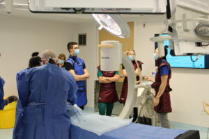
Throughout the three-hour lab, Desmond and the students stepped in periodically to capture images of the ankle cadaver, which immediately appeared on a screen for the surgical team to assess. All three of the students expressed what a fantastic opportunity this was for them to get more hands-on experience using the medical imaging equipment.
“At my first clinical placement, we didn’t have a lot of opportunity to see the operating room, so I found it really interesting to see how different professions worked together and especially how my profession ties into everyone else’s,” says Stephanie Olson.
“Michener strives to provide our students with the most useful and contemporary experiential learning opportunities and we are always looking for opportunities to use our impressive facilities to support these learning experiences,” says Harvey Weingarten, Principal of Michener. “This lab is but one example.”