Sonographers’ Role in Medical Diagnosis
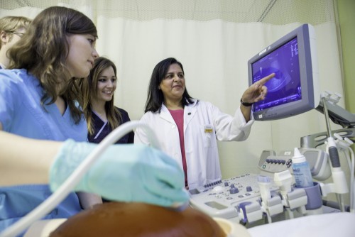
 Diagnostic Medical Sonography, more commonly known as ultrasound, is a rapidly changing technology that is used by Sonographers to assess and help diagnose medical conditions. “Sonographers use ultrasound technology as well as their knowledge, skills and aptitude to look for abnormalities or pathologies in the human body,” says Sheena Bhimji Hewitt, Ultrasound Professor at The Michener Institute and Chair of Sonography Canada Board. She explains that both academic and clinical education is paramount to the creation of a competent and safe Sonographer. “When the Sonographer is competent and able to recognize normal from abnormal findings, they are ensuring the patient’s prognosis and wellbeing.”
A Diagnostic Tool to Improve Patient Outcomes
Sonographers, working in hospitals, private clinics, research facilities, or other healthcare environments, display competency in their academic knowledge, communication and collaboration skills in order to provide safe and high quality patient care. Sonographers must not only be able to write comprehensive reports, but also have the skills to assess the image and follow up appropriately.
As ultrasound becomes increasingly accessible and portable, the dynamic, real-time nature of this technology allows trained professionals to provide point-of-care assessment for patients undergoing interventional, emergency procedures or examinations. “Sonography is one of the best real time diagnostic technologies medical professionals have,” says Sheena.
What Disciplines Can The Sonographer Specialize In?
Although ultrasound technology is typically associated with pregnancies, there are actually three main disciplines of sonography:
- A Generalist specializes in abdomen, gynecological, superficial structures (such as the breast, thyroid or scrotum) as well as obstetric exams (pregnancy to delivery).
- A Vascular Sonographer is an expert in assessing the anatomy and physiology of blood vessels on the adult population.
- An Echocardiographer specializes in assessing the heart as well as the major vessels associated, and can specialize in either pediatric or adult sonography.
What About 3D/4D Ultrasound? What Do Advances In Technology Mean For Patient Care?
Recent developments in technology such as 3D/4D ultrasound are mostly used during pregnancy ultrasounds to display 3-dimensional images and movement of the fetus, allowing for a better external view.
Ultrasound is also being integrated into other diagnostic and therapeutic medicines. For example, it is used in targeting, localizing and verifying prostate, gynecological and breast cancers in image guided radiation therapy (IGRT). “Sonography is regarded as a technology that is non-invasive and versatile. The future for it is limitless,” says Sheena. “In the past ten years, ultrasound technology has expanded into areas such as musculoskeletal ultrasound in sports medicine and can be used by rheumatologists for arthritis studies.”
U ltrasound Is for You If you…
- Want a challenging career that requires a very high level of knowledge, skills and responsibility
- Are physically fit and have excellent hand eye coordination and spatial reasoning skills
- Can multi task, problem solve, critically analyze, be organized and efficient
- Have effective communication and collaboration skills and can handle stressful situations
- Can interact with patients in an empathetic and non-judgmental manner and believe in quality and safe patient centered care
Want to become a Sonographer? Here’s how:
At The Michener Institute, the Ultrasound Program curriculum is designed using the Sonography Canada National Competency Profile (NCP) and is accredited by the Canadian Medical Association. A graduate from the Michener Ultrasound Program is prepared to write the Sonography Canada credentialing exams (Clinical and Academic exams) to become a Generalist Sonographer. The graduate’s competency is validated with the achievement of the CRGS (Canadian Registered Generalist Sonographer) credential.
Fun Fact about Diagnostic Medical Sonography:
Commander Chris Hadfield, Canadian Astronaut, and his team performed vascular ultrasounds so that experts could evaluate changes and effects of the body in zero gravity!
Ultrasound Lab – the rig we used to measure how Tom’s heart is changing and adapting without gravity, like rapid aging. pic.twitter.com/9MMG3XEr
— Chris Hadfield (@Cmdr_Hadfield) January 1, 2013
 ltrasound Is for You If you…